EV Array
High-throughput Multiplexed Phenotyping of Extracellular
Vesicles (EVs)
Extracellular Vesicles
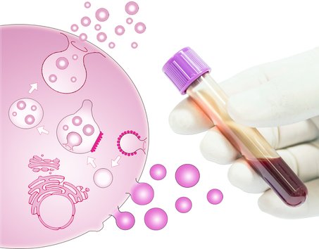
All living cells produce extracellular vesicles (EVs), which are NANO-SIZED CELL-DERIVED COMPARTMENTS. They are considered as a pivotal part of the intercellular environment and act as important players in cell-to-cell communication.
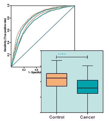
The fact, that EVs are involved in the development and progression of several diseases, has formed the basis for the use of EV analyses in a CLINICAL SETTING and envisions a great potential for using EVs as DISEASE-RELATED BIOMARKERS.
EVs are a heterogeneous population of vesicles that can be divided into a number of subpopulations based on specific characteristics such as size, biogenesis, cellular origin, protein composition, and biological function. The two major subtypes of EVs are exosomes and microvesicles.
Development and standardization of rapid and inexpensive methods would pave the way for widespread use of extracellular vesicle-based cancer diagnosis in both developed and developing countries.
- Choi et al., Mol Diagn Ther (2013) -
Principle of the Extracelluar Vesicle Array
EV Array
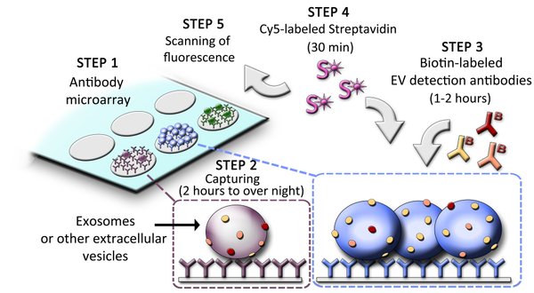
Step 1: The EV Array is composed of different capture antibodies printed on a microarray slide.
Step 2: 10-100 µL plasma or other body fluids (urine, saliva, BALF, etc.) are applied in a 96-well setup and incubated 2 hours to overnight.
Step 3: The EVs are detected with a cocktail of biotinylated antibodies.
Step 4/5: The presence and thereby phenotype of EVs is visualized after incubation with Cy5-labeled Streptavidin using a fluorescence scanner.
The use of microarray as a platform with spots of 1 nL volumes minimizes the cost of the EV Array as only small amounts of antibodies are needed.
The EV Array has been tested and optimized for various bodyfluids:
Body Fluid | Optimal Volume |
Plasma | 10 µL |
Urine | 50 µL |
Saliva | 100 µL |
Synovial Fluid | 50 µL |
Cerebrospinal Fluid | 50 µL |
Ascites | 10-20 µL |
Bone marrow | 10 µL |
Bronchoalveolar Fluid | 50 µL |
Cell supernatants | 100 µL |
Advantages of the EV Array
The research field of EVs has shown a great demand for commercial available technologies to analyse EVs for biomarkers. Currently, extensive and time consuming (> 24 hours) purification procedures is needed prior to analysis of single markers. NO OTHER existing technology phenotypes EVs in a multiplexed manner using unpurified material.
The EV Array consumes ONLY 10 – 100 µL SAMPLE, whereas purification procedures uses several milliliters. The EV Array is performed in multi-well cassettes in a high-throughput manner (up to 21 samples per slide), but is still easy to handle in the laboratory.
The use of microarray as a platform with spots of 1 nL volumes minimizes the cost of the EV Array as only small amounts of antibodies are needed.


